
 Skin lumps (or masses) are very commonly seen in dogs but are rarer in cats. Occasionally you will be able to feel a lump under the skin (such as a lymph node) and rarely within the abdomen.
Any new lump should always be investigated, particularly if it appears suddenly and is growing.
The first thing we will try to establish in consult is whether the mass is inflammatory, infectious or a neoplastic (cancerous) mass. Once we determine that a mass is cancerous we then need to work out if it is malignant (will spread and grow quickly) or benign (slow to spread and slow growing.)

In the consultation we will first visually examine the lump and assess the surrounding skin, check the local lymph nodes and perform a full physical examination. Some common inflammatory conditions are allergic hives or bacterial dermatitis. In these cases we will prescribe medication and the skin lesions (lumps) usually resolve.


In cats, abscesses are common in outdoor cats and are caused by wounds from fighting and these appear suddenly and are painful to touch. They often require surgical lancing to clear the infection, then ongoing medication to control infection and inflammation.
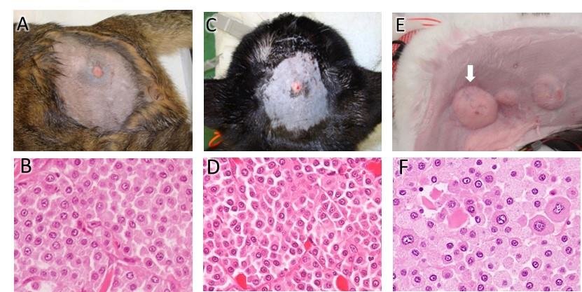
 If we cannot confirm an infection or inflammation by looking alone, the next step is to obtain a tiny sample of cells from the lump by performing a “fine needle aspirate”. This involves inserting a small needle into the lump and extracting the cells, then examining these under a microscope (called cytology). This procedure is usually well tolerated by our patients without the need for sedation but it depends on the location of the mass and the temperament of the patient.
Again the first step when performing cytology is to differentiate inflammation from neoplasia and we can often tell this by the type of cells we see under the microscope. We can also get clues sometimes as to whether the mass is likely to be benign and less of a concern or if it is a type of lump that can be malignant (more aggressive type of cancer).
For a deeper mass (such as an abdominal mass) we will often need to perform an ultrasound as the first diagnostic step.
When we have more information on the lump we can come up with a plan to consider surgical removal. Generally it is always a good idea to remove any rapidly growing mass and then send it off to our dedicated veterinary laboratory for further testing (histopathology) which will then give us a definitive (final) diagnosis.
There may be cases when surgery is not warranted – for example on a geriatric patient with multiple benign lumps (such as warty papillomas or fatty lipomas) where the risk of surgery outweighs the benefit of removing the mass/es. On the whole though we would usually recommend surgery as the best way of dealing with any new mass, particularly if we are not sure what we are dealing with.
 In the event of suspected malignant mass we will have to plan our surgery to ensure we get enough tissue around the lump so it doesn’t regrow and further tests may be suggested. We usually perform a blood test but may also require chest X rays (to check for metastasis – spread of cancer), examination of the lymph nodes and sometimes an ultrasound of the abdomen.
 So, if you do find a new lump on your pet, please bring them in for a consultation as soon as possible. You may end up relieved and they may only require some medication or we may plan to remove the lump to reduce the risk of spreading cancer. Either way, it is best to get onto it early as we know with any type of lump, the earlier we catch it, the better the prognosis and chance of a quick recovery.